

Asking a metabolic question inside a human tumor
Reading time: about 6 minutes. Nothing here is medical advice.
The container is smaller than you expect. It travels a few hundred yards, from an operating room to a bench, in a cooler that could just as easily hold a sandwich. For the hour or so that follows, the cells inside are still alive. They do not know the tumor they came from has been removed. They go on doing what they were doing.
That last sentence is the reason for a great deal of what my lab does. They go on doing what they were doing — and what they were doing is something we have historically measured by what those cells look like, rather than by what they are running on.
A cell can be present and still be useless
The immune system can find a tumor. This is not the difficulty. In head and neck cancers, as in most solid tumors, you can take a piece of tissue and count a great many T cells inside it — cells whose entire biological purpose is to recognize something abnormal and destroy it.
They are there, and the tumor is also there.
The word the field uses for this is exhaustion: a state in which a T cell that has been stimulated for too long, too continuously, stops responding. It is not death, and it is not absence. The cell persists, keeps its identity, and stops working. Checkpoint blockade — the class of drugs that includes anti–PD-1 antibodies, and the reason immunotherapy entered common vocabulary — was built to release one of the brakes holding those cells back. For some patients it works remarkably well. For many it does not, and the honest answer to why is still incomplete. Baessler and Vignali’s 2024 review in Annual Review of Immunology is the best current account of what exhaustion is and how it is programmed; Chow and colleagues have written the clinical counterpart, which argues something worth sitting with — that exhaustion may be as much a reflection of poor tumor control as a cause of it.
Why what a cell burns might matter
Here is an idea borrowed from an older part of immunology, one that has been steadily gaining ground: a cell’s capacity to do work is constrained by how it makes energy.
This is less abstract than it sounds. A T cell that has just recognized its target and is preparing to divide and kill has enormous, immediate demands, and it meets them differently from a cell that is sitting quietly and expecting to persist for years. Different pathways, different fuels, different consequences for what the cell can physically do next. Metabolism is not a housekeeping detail underneath the interesting biology. In a T cell it is close to being the interesting biology.
This is not speculation. Effector T cells preparing to divide and kill run heavily on aerobic glycolysis; memory T cells, built to persist, favor fatty acid oxidation and oxidative phosphorylation — and Buck and colleagues showed in 2016 that the shape of a cell’s mitochondria is itself part of what enforces the difference. Metabolic Instruction of Immunity is the review I would hand anyone starting here.
If that is right, then a cell’s metabolic state is not merely a readout of how it is doing. It is part of the reason it can or cannot do anything. And unlike a fate that has already been sealed, metabolism is the kind of thing you can, in principle, interfere with.
That is the possibility worth taking seriously: not to add another brake-release, but to change what the cell has available to work with.
The measurement problem
There is a reason this has been studied more thoroughly in mouse spleens than in human tumors.
The classical way to measure what a cell is running on is to take a great many cells, put them in an instrument, and watch how they consume oxygen or acidify their medium. It is a good method and it answers a real question. It also requires a large, pure population, and it destroys the sample.
A human tumor gives you neither luxury. What arrives in that small cooler is a mixed and finite thing — tumor cells, stroma, several immune lineages, some of them present in the low thousands. The population you most want to ask about is frequently the rarest one there. By the time you have sorted enough of it to fill an instrument, you have used the sample and learned about an average.
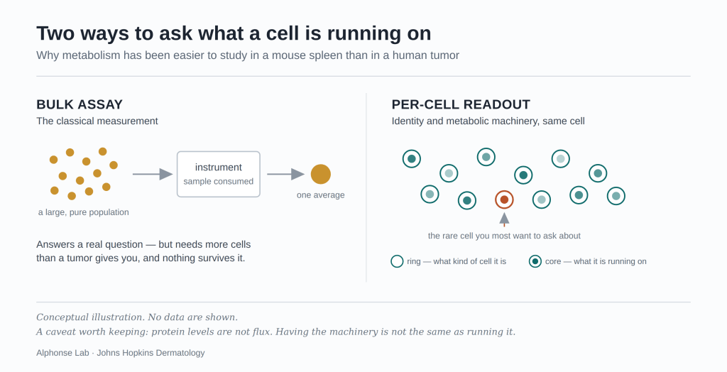
The workaround is to measure metabolic machinery inside individual cells, at the same time as everything else about them — which enzymes and transporters a cell has invested in, alongside the surface markers that say what kind of cell it is and what state it is in. High-parameter spectral flow cytometry makes this possible: enough simultaneous measurements per cell that identity, activation state and metabolic program can be read off the same cell rather than inferred across separate experiments.
It is a compromise. Protein levels are not flux; having the machinery is not the same as running it, and I would not want that elided. But it lets you ask the question in the tissue where it matters, in the cells that are actually rare, one cell at a time.
The window a neoadjuvant trial opens
There is a second reason to ask this in head and neck cancer, and it has less to do with biology than with trial design.
In a neoadjuvant trial, treatment is given before surgery rather than after. The clinical rationale is its own subject. The consequence for laboratory work is rare: tumor tissue from the same patient before treatment and after it. Not a model. Not a cell line. The same person’s disease, twice, with a defined intervention in between.
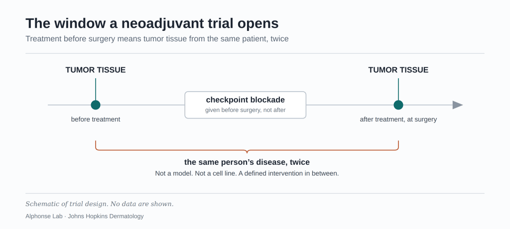
Correlative work on specimens from trials of this design is where our questions have been aimed.
Two markers guided where we looked, both well described by others. CD39 is an ectoenzyme carried by chronically stimulated T cells — and, in a finding that complicates any simple reading of it, Duhen and colleagues showed that CD39, together with CD103, marks the CD8 T cells in solid tumors that are actually tumor-reactive, with an exhausted tissue-resident phenotype. In head and neck cancer specifically, more of those cells went with better overall survival. OX40 sits on the other side of the ledger: a costimulatory receptor of the TNF receptor superfamily that supplies the second signal a T cell needs to activate fully. Neither is our discovery. The question we brought to them was narrower: whether cells carrying these markers differ in what they are running on, and whether that difference is the kind of thing anyone could act on.
What I do not yet know
It would be dishonest to write this without the list, so here it is.
Does metabolic state cause dysfunction, or report it? In an observational study, a cell that has stopped working and a cell with an altered metabolic program are the same cell. Establishing direction requires perturbation, and perturbation requires material that human tumor studies rarely provide.
Does a cell in a dish still resemble the cell that was in the tumor? Some of this work necessarily happens after the cells have left the tissue. That gap is real and I do not think it can be argued away — only bounded, and stated.
Can you change metabolism in one population without breaking another? Metabolic pathways are not specific to lineages. Anything that alters fuel use in an exhausted T cell will also reach cells you would rather leave alone. Whether a useful window exists is an open question, not a solved one.
Coda
This piece is deliberately about the question and not the answer. I will come back to what we found.
The container comes up from the operating room. Someone signs for it. For an hour, cells that spent months inside a tumor are still alive on a bench, still doing whatever it was they had learned to do there — and for that hour, it is possible to ask them what they were running on.
Not much of a window. But it is the one we have, and it opens onto a person rather than a model.
References
Baessler A, Vignali DAA. T cell exhaustion. Annu Rev Immunol 2024;42:179–206. doi:10.1146/annurev-immunol-090222-110914
Chow A, Perica K, Klebanoff CA, Wolchok JD. Clinical implications of T cell exhaustion for cancer immunotherapy. Nat Rev Clin Oncol 2022;19:775–790. doi:10.1038/s41571-022-00689-z
Buck MD, O’Sullivan D, Klein Geltink RI, et al. Mitochondrial dynamics controls T cell fate through metabolic programming. Cell 2016;166:63–76. doi:10.1016/j.cell.2016.05.035
Buck MD, Sowell RT, Kaech SM, Pearce EL. Metabolic instruction of immunity. Cell 2017;169:570–586. doi:10.1016/j.cell.2017.04.004
Duhen T, Duhen R, Montler R, et al. Co-expression of CD39 and CD103 identifies tumor-reactive CD8 T cells in human solid tumors. Nat Commun 2018;9:2724. doi:10.1038/s41467-018-05072-0
Buchan SL, Rogel A, Al-Shamkhani A. The immunobiology of CD27 and OX40 and their potential as targets for cancer immunotherapy. Blood 2018;131:39–48. doi:10.1182/blood-2017-07-741025
Martin P. Alphonse is an Assistant Professor in the Department of Dermatology at Johns Hopkins University School of Medicine. Views here are his own.

