
What we found when we asked a metabolic question inside a human tumor
Reading time: about 7 minutes. Nothing here is medical advice.
In an earlier post I described a small cooler carried from an operating room to a laboratory bench, and the hour afterward in which the cells inside are still alive — still doing whatever they had learned to do inside a tumor. I ended it with a promise: that I would come back to what we found.
The paper was published this week in Cancer Immunology Research, and Johns Hopkins Medicine released a news story on the work the following day. This is an account of what it shows, and — at the same length — of what it does not.
What we set out to measure
A short reprise for anyone arriving here first. In head and neck cancer, as in most solid tumors, the immune system finds the tumor. T cells are present in large numbers. Many of them are exhausted: still there, still themselves, and no longer working. Checkpoint blockade — anti–PD-1 antibodies — releases one of the brakes on those cells, and for a minority of patients it works well.
We wanted to know whether those cells differ in what they are running on, and whether any such difference is the kind of thing that could be acted on. To ask it, we measured metabolic enzymes inside individual cells at the same time as the markers that say what each cell is, using high-parameter spectral flow cytometry on tumor tissue from patients in two neoadjuvant trials at Johns Hopkins. One trial gave anti–PD-1 alone before surgery. The other combined it with an antibody against interleukin-8 (IL-8), a signal thought to promote suppression by myeloid cells in tumors.
Who the study is about
Before any finding, the population — because it bounds everything that follows.
The study drew on 15 patients and 25 tumor specimens. Where a tumor yielded both a core and a peripheral sample we kept them separate, because the biology of the two regions differs. This is a mechanistic study, not a clinical one, and I would not want anything that follows read otherwise.
Four findings
One immune population expanded — under one treatment. Mucosal-associated invariant T cells — MAIT cells, an unconventional T-cell lineage that recognizes small molecules made by microbes rather than conventional peptide fragments — increased in tumors after anti–PD-1 combined with anti–IL-8, but not after anti–PD-1 alone. We tested that difference between the two trials directly, rather than inferring it from one result being significant and the other not. Conventional CD8 T cells and innate lymphoid cells showed no detectable change, although with 12 or 13 specimens per timepoint we could not have detected modest ones.
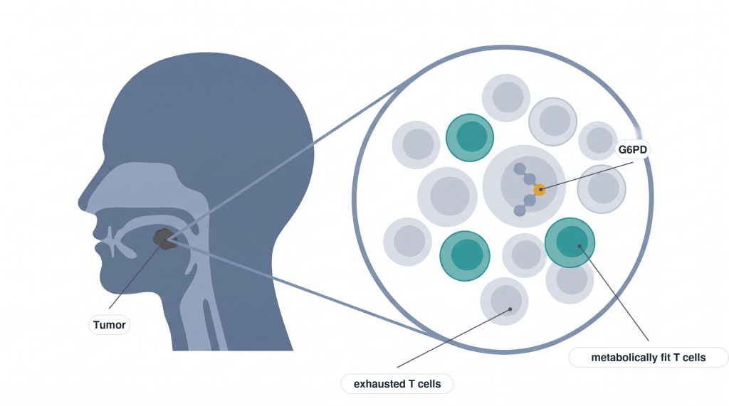
Some T cells look metabolically fit, and they carry a recognizable marker. Across MAIT cells and conventional CD8 T cells, those lacking CD39 but carrying OX40 — the two markers from the earlier post — had a profile we read as metabolically fit and less exhausted: more of the machinery associated with oxidative, energy-generating metabolism, and less of the machinery for building biomass. I want to be exact about this. It describes what those cells are. It does not show that they increased with treatment, and it does not show that they predict who will respond; our cohort is too small and too uniformly non-responding to say either.
The exhausted cells carry more of one particular enzyme. T cells lacking OX40 had more G6PD — glucose-6-phosphate dehydrogenase, the rate-limiting enzyme of the pentose phosphate pathway, a branch of glucose metabolism. That makes the pathway a candidate target: a place where one might intervene. The caveat from the earlier post applies with full force. We measured the enzyme, not the pathway’s activity. Having the machinery is not the same as running it.
In the laboratory, blocking that enzyme changed how exhausted cells responded to anti–PD-1. Using CD8 T cells from untreated patients, separated by how much CD39 they carried, we added anti–PD-1, a G6PD inhibitor, or both, in culture. Cells at the three levels of CD39 responded differently. In the most exhausted cells, only the combination turned down genes of the STAT3 and mTOR pathways, and it increased release of soluble CD27, a sign of T-cell activation.
This was tested only in the laboratory, in cells taken from patient samples — not in patients. No patient received the metabolic drug.
And one population that may stand in the way
We also looked at innate lymphoid cells, a lineage without antigen receptors that shapes the tissue environment early in an immune response. A subset of type 2 innate lymphoid cells, marked by high levels of CD294, had an immunosuppressive profile and carried high levels of two proteins, CCR8 and CPT1A. We propose that these cells may limit how well checkpoint blockade works.
Coda
The container still comes up from the operating room. What has changed is that we know a little more about what to ask the cells inside it — which ones look ready to work, which enzyme the exhausted ones carry, and which neighbors may be holding them back.
That is not an answer. It is a better question.
Immunometabolic profiling of T cells and innate lymphoid cells uncovers therapeutic targets to enhance immunotherapy in head and neck cancer. Cancer Immunol Res 2026. https://doi.org/10.1158/2326-6066.CIR-26-0245
Johns Hopkins Medicine’s account of the work, published 30 September 2026: Johns Hopkins investigators identify a metabolic weak point in exhausted immune cells in head and neck cancer.
Martin P. Alphonse is an Assistant Professor in the Department of Dermatology at Johns Hopkins University School of Medicine. Views here are his own.

